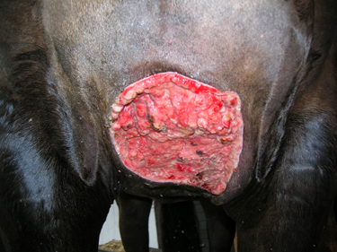
Hendrix is a ten year old bay gelding that presented in May 2006 with a sarcoid on his chest. Initially the sarcoid had a fleshy, ulcerated appearance with a large fluid pocket behind it. Over the following two weeks the swelling on his chest increased in size and was subsequently lanced. Two buckets of pus were drained from the area and several sarcoid nodules were shelled out.
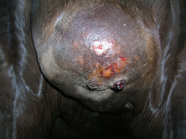
There are many different types of sarcoids and therefore their appearance can differ greatly. Some are hairless areas of crusting (occult), wart-like (verrucose), nodular, fleshy, ulcerated, bleeding (fibroblastic) or mixed. They are generally considered to be a type of ‘skin cancer’ which is limited to the skin and underlying tissue. Sarcoids do not ‘spread’ to internal organs though they can be locally aggressive. They characteristically are unpredictable and therefore can be difficult to treat.
Hendrix was referred to Liverpool University in June 2006 for laser surgery, which was performed following 10 days of antibiotic therapy to control infection and reduce the swelling. He remained at the hospital for almost 4 weeks before returning home. When at home, his rider had to flush his chest wound daily with 5 litres of salt water, he was kept on box rest and walked out in hand daily for a further 3 weeks.
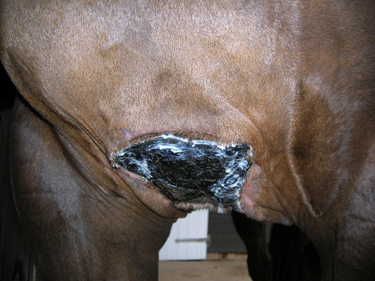
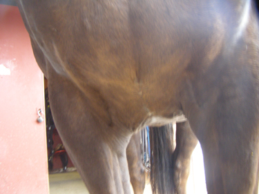
In August 2006 excess proud flesh (granulation tissue) was trimmed from the wound margins and sent to Professor Derek Knottenbelt at Liverpool university for histopathological analysis. This confirmed that there were still sarcoid cells present and we were given a treatment protocol for AW4 (’sarcoid cream’) applications. The sarcoid cream resulted in a hard plaque being formed which took 2 months to slough off.